Professor H. Peter Soyer and Katie Lee have published a review article in the International Journal of Dermatology and Venereology.
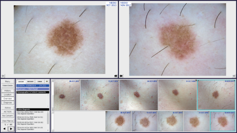
The article reviews recent developments in teledermoscopy and total body photography in the diagnosis and monitoring of skin lesions, including skin cancers and other inflammatory lesions.
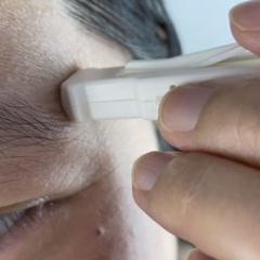
It gives an overview of current practices in mobile teledermoscopy and total body dermoscopy.
It also looks at the role of artificial intelligence as an analytical tool to help decision making for clinicians in the future.
Lee, KJ & Soyer, HP. (2019) Future developments in teledermoscopy and total body photography. Int J Dermatol Venereol, 2019,2(1):15-18. doi: 10.3760/cma.j.issn.2096-5540.2019.01.003