 According to a position statement issued by the European Society of Radiology (ESR), “Medical imaging has always been personalised and in the future it will prove fundamental to almost all aspects of personalised medicine” [1].
Professor Hedvig Hricak, Chair of the Department of Radiology at Memorial Sloan-Kettering Cancer Center in New York, even stated in 2010 that medical imaging had become “a ‘guiding hand’ of personalized medicine for cancer care” [2]. In early diagnosis and treatment monitoring of melanoma and skin cancer, especially, medical imaging has always played a key role.
However, there is much more to come, as Professor H. Peter Soyer, The University of Queensland, Australia, pointed out at the EADV Congress in Geneva today. “Nowadays, new high-tech imaging, combined with artificial intelligence and decision support systems will surely redefine the early diagnosis of melanoma and skin cancer!”
A landmark paper was published this February in the internationally renowned journal “Nature” [3]. The authors tested a machine learning, convolutional neural network (CNN) for the classification of skin cancer. CNNs are feed-forward artificial neural networks and learn the filters that in traditional algorithms were hand-engineered. This makes them independent of prior knowledge and human effort. The working group at Stanford University in California used only pixels and disease labels as inputs, the system being “fed”, or trained, with a dataset of 129,450 clinical images consisting of 2,032 different diseases (two orders of magnitude larger than previous datasets). The CNN was then tested against 21 board-certified dermatologists on biopsy-proven clinical images, who firstly had to identify the most common skin cancers, and secondly the deadliest skin cancer. The result was astonishing: the CNN achieved a performance on par with all tested experts across both tasks, demonstrating an artificial intelligence capable of classifying skin cancer/melanoma with a level of competence comparable to dermatologists. It was able to classify skin cancers/melanomas as “biopsy/treatment is needed” or “reassure the patient/everything is fine” as reliably as the experts. According to the authors of the study, deep neural networks deployed on mobile devices can potentially extend the reach of dermatologists outside the clinic. They believe that this technique “can potentially provide low-cost universal access to vital diagnostic care” [3]. The question is, can this really become true – or is it just a “Silicon Valley dream”?
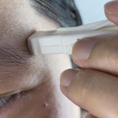
As Professor Soyer explained in Geneva, this development of artificial intelligence and its adoption in dermatology is proceeding rapidly, indeed, and the market for AI technologies is flourishing. One area of focus is on automated analysis of features in dermoscopic images of skin lesions (shape, colour, size), another on identifying potential characteristics of melanoma (i.e. border irregularity, multiple colours within lesions), and AI is being integrated into software for different types of imaging platforms (3D imaging systems, digital dermoscopic images and smartphone dermoscopic images). He also believes that smartphone dermoscopic imaging with built-in AI is likely to be the most accessible method for skin lesion analysis in future.
There are pitfalls, however, and dermatologists should seek to ensure minimum quality standards for such devices. One study of current smartphone apps [4] for diagnosing melanoma showed that the app market is a fast-moving one, with almost half of 2014 apps no longer existing in 2017. At present, 10 melanoma apps have integrated image analysis functions, but their accuracy varies considerably. When three apps with AI were tested on skin lesions in the clinical setting in 2016, sensitivity was found to range from 21 to 72% and specificity from 27 to 100%. “As technology becomes more commonplace in dermatological practice, it is essential to review continuously the accuracy of these devices”, explained Professor Soyer. “The evidence base of many of these apps is improving, but it remains limited due to the vast majority of apps not being peer-reviewed.” According to Soyer, the apps available should be certificated by dermatologists. “We have to ensure a minimum rate of false-positive and false-negative diagnoses. In the end, we are talking about melanoma, a fatal disease that people die of.”
Soyer emphasised that these techniques have definite advantages. One study [5] has shown that they consistently reduced waiting times to assessment and diagnosis, and that patient satisfaction levels were high. “These devices will change the day-to-day practice of dermatologists. We will more or less exclusively see patients with cancer and suspicious lesions that have to be removed. As a consequence, we will have more time for counselling on treatment, etc., because we will not have to see all the patients who have harmless skin lesions.”
Asked whether artificial intelligence can diagnose skin cancer/melanoma more reliably than dermatologists in the future, Soyer gives an ambiguous answer: “Technically, yes, because AI systems can be fed with more images than an experienced real-life dermatologist can see in his or her lifetime. But it cannot process contextual information, like family anamnesis or other symptoms. It doesn’t see the whole patient. For these reasons, I am quite optimistic that human dermatologists will always be needed!”
Media: Professor H. Peter Soyer, p.soyer@uq.edu.au
[1] European Society of Radiology (ESR). Medical imaging in personalised medicine: a white paper of the research committee of the European Society of Radiology (ESR). Insights Imaging 2015 Apr; 6(2): 141-55.
[2] European Society of Radiology. Medical imaging in personalised medicine: a white paper of the research committee of the European Society of Radiology (ESR). Insights Imaging 2011 Dec; 2(6) :621-630.
[3] Esteva A, Kuprel B, Novoa RA et al. Dermatologist-level classification of skin cancer with deep neural networks. Nature. 2017 Feb 2; 542(7639): 115-118
[4] Ngoo A, Finnane A, McMeniman E et al. Efficacy of smartphone applications in high-risk pigmented lesions. Australas J Dermatol. 2017 Feb 27. doi: 10.1111/ajd.12599. [Epub ahead of print]
[5] Finnane A, Dallest K, Janda M, Soyer HP. Teledermatology for the Diagnosis and Management of Skin Cancer: A Systematic Review. JAMA Dermatol 2017 Mar 1; 153(3): 319-327.