Dermoscopy has been a key tool in the advancing methods for early detection of skin cancers and melanomas for more than 30 years.
Professor H. Peter Soyer, Chair in Dermatology and Director of the Dermatology Research Centre, The University of Queensland, is considered a pioneer and world leader in the field of dermoscopy.
We take the opportunity to ask Professor Soyer about his career using dermoscopy, his views on its place in the future, and what it means have published the Dermoscopy text book in its third edition.
Tell us about when you first started dermoscopy?
We first started to use dermoscopy (then called surface microscopy) in 1984, when I was a registrar at the Dermatology Department at the University of Graz, in Graz, Austria. Then in 1989, when I was an attending physician, recently awarded Universitaets-Dozent (traditional PhD equivalent in the German speaking world), I held my first dermatoscope. We were fortunate to get it first hand as it was developed by our colleagues at the Munich Department of Dermatology and Heine. I recall how excited we all were to use it for the benefit of our patients and for our research projects. Since then I have used dermoscopy in countless patients and was involved with numerous educational activities around the world.
How do you use dermoscopy in your practice now?
Since that time, with the development of many more dermatoscopes I have used dermoscopy constantly in clinical practice. Yes, I have used naked eye examinations, but I have used dermoscopy for almost every lesion, particularly the banal lesions. This is so important as I feel you must train your eye with the clear cut lesions to get an overview of the biologic ecosystem of moles, seborrhoeic keratoses and other lesions.
What is the future of dermoscopy?
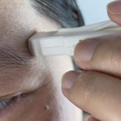
It is an integral part of the clinical examination. Just as you need a good light source, you need a good lens. It’s critical to use a dermatoscope.
Digital dermoscopy has changed the workflow, with all the photographic documentation. We are now at the beginning of artificial intelligence, and the issue is to which extent new imaging technologies will produce pseudo-dermoscopy images without a dermatoscope. Of course this will not come straight away, but I expect in the years to come, we will have dermoscopic quality images, without using a dermatoscope.
What is your advice to doctors who are starting to learn dermoscopy?
I would say, just start using it. Use it for lesions you know the diagnosis for. This will give you a threshold for what you know. When you know the features of classic lesions, then you can begin to work on the more complex ones.
What does it mean to have Dermoscopy: The Essentials published in the third edition?
We published the first edition of Dermoscopy: The Essentials in 2004, and have continued to update and evolve with the technique over the following years. It is exciting to be able to see a new generation of dermatologists and general physicians becoming dermoscopy experts.
Dermoscopy: The Essentials, Third Edition was published by Elsevier, 2020.
Soyer, H. Peter, Argenziano, Giuseppe, Hofmann-Wellenhof, Rainer and Zalaudek, Iris (2020). Dermoscopy: The Essentials. 3 ed. Edinburgh, Scotland, United Kingdom: Elsevier/Saunders.